Science and emerging technology
While Patients Wait
The AI that reads a brain MRI in seconds is here. Deploying it where it matters is another question.
Designed for the person who isn’t there · 4 of 10 essays in this pattern
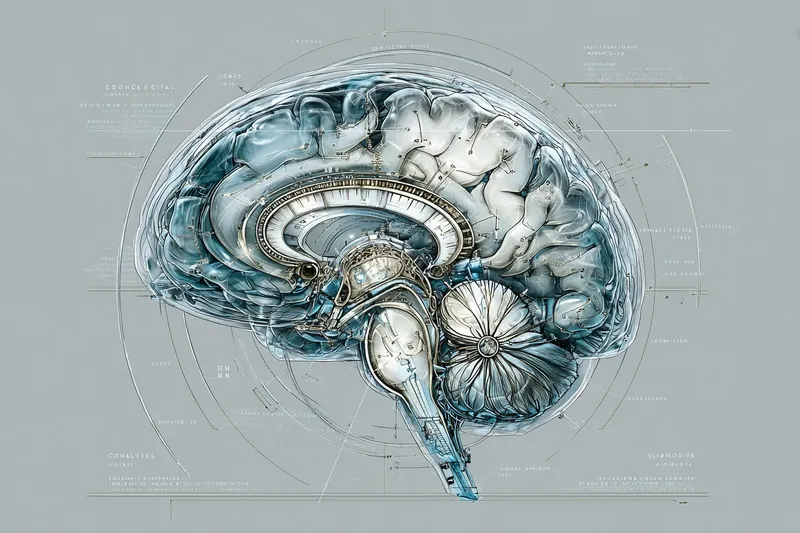
A case documented in the medical literature described a patient with pancreatic cancer who missed out on a potentially life-prolonging treatment because their scan result was delayed by eighteen months. The scan had been taken. The images existed. No one had looked at them in time.
That case was not unusual enough to be a scandal. It was unusual enough to make the journal. The background against which it sits — a radiology system under sustained and worsening pressure, in which delays are the norm rather than the exception — has become so familiar that it no longer registers as news. It is simply the condition patients navigate when something goes wrong inside their heads and they need to know what it is.
In September 2025, more than 386,000 people in England were waiting longer than six weeks for a diagnostic test. The total number waiting for any diagnostic test exceeded 1.7 million. The Royal College of Radiologists has documented a 30% shortfall of clinical radiologists in England — roughly 2,000 doctors short of what is needed to meet current demand — with the gap projected to reach 39% by 2029. Forty-six percent of NHS acute trusts are not meeting even the interim waiting time target.
A system that can read a brain MRI scan in seconds has arrived. Not a prototype. Not a laboratory demonstration. A system trained on more than 220,000 real clinical scans, tested across more than 30,000 studies in a running health system over a full year, published in February 2026 in Nature Biomedical Engineering. The question is not whether the technology works. It is why, given that it works, the gap between scan and diagnosis remains so wide for so many patients.
What the AI can actually do
The system is called Prima. It was developed by a team at the University of Michigan led by neurosurgeon Todd Hollon, and it works differently from most AI diagnostic tools that have preceded it.
Earlier AI models in radiology were built for specific tasks: detecting a particular kind of lesion, flagging a certain cancer type, screening for a defined abnormality. They were useful for the thing they were designed for and nothing else. Prima was trained on everything — every MRI study taken at the University of Michigan Health system since radiology records were digitised, covering more than 220,000 studies and 5.6 million imaging sequences. It was trained not just on the images themselves but on the patients’ clinical histories and the reasons each scan had been ordered by a physician. It works, as its developers describe it, the way a radiologist works: integrating what the image shows with what the patient’s medical history tells you.
Across more than 50 different radiological diagnoses — stroke, brain haemorrhage, tumours, multiple sclerosis, aneurysms and more — Prima outperformed other state-of-the-art AI models, achieving a mean diagnostic accuracy of 92% across that range. It can identify which cases require urgent attention and immediately alert the appropriate specialist: a stroke neurologist if the scan suggests a stroke, a neurosurgeon if it suggests a bleed. Feedback is available as soon as imaging is complete. The result that would otherwise take days or weeks to arrive takes seconds.
Prima was trained on every MRI taken at a major health system since records were digitised. It works the way a radiologist works — integrating image data with clinical history. The result takes seconds.
UCL and King’s College London published parallel work in December 2025. Their model, trained on 60,000 brain MRI scans and the radiology reports written about them, accurately distinguished normal from abnormal scans approximately 19 times out of 20. A randomised multicentre trial across UK hospitals started in 2026 to assess how the system performs in live clinical workflows. These are not the same research programme, but they point in the same direction: AI that can read a brain scan fast and flag what matters is operationally real, not theoretical.
For acute neurological conditions, stroke above all, the stakes of speed are measurable in the specific. In stroke care, the standard principle is time is brain: for every hour of untreated ischaemic stroke, roughly 120 million neurons are lost. The gap between scan and treatment decision is a gap in which irreversible damage accumulates. A system that compresses that gap from hours to seconds is not a marginal improvement in workflow efficiency. It is a different category of clinical event.
Why the gap persists
If the technology exists and the problem is severe, the natural question is why the gap between them remains so large. The answer is not simple and it is not flattering to any single part of the system.
Regulatory approval is one obstacle. AI diagnostic tools that make clinical decisions about individual patients are classified as medical devices in the UK and the EU. The approval process is rigorous — necessarily so, given the consequences of getting it wrong — but it is not designed for the pace at which AI research is producing results. The models that receive approval tend to be narrow-task systems, because narrow tasks are easier to validate; a foundation model like Prima, which makes diagnoses across 50 conditions, presents a different and more complex challenge. The UCL team’s next step — a randomised multicentre trial in UK hospitals — is the appropriate pathway, and it is one that takes years.
Integration is a second obstacle. An AI model that produces a result in seconds is only useful if that result reaches the clinician who needs it, in a format they can act on, connected to the patient record system they are already using. NHS radiology departments typically operate on legacy IT infrastructure. The connections between imaging systems, electronic patient records, and clinical communication tools are incomplete, inconsistent, and often not interoperable. The radiologist shortage means that even when AI flags a case as urgent, there may not be a specialist immediately available to review the flag and make the treatment decision.
Liability sits beneath both. When an AI system incorrectly categorises a scan — flags something urgent that is not, or misses something that is — the question of who is responsible is unresolved in most legal and professional frameworks. The EU AI Act, whose main provisions came into application in August 2026, classifies medical AI diagnostic tools as high-risk and requires documentation of training data, bias checks, and human oversight policies — though the provisions specifically covering CE-marked medical devices are not due to apply until 2027. Radiologists operating under professional accountability for their diagnoses have every reason to be cautious about delegating to a system whose error profile is not yet fully understood.
The technology compresses the gap from hours to seconds. The regulatory pathway, the IT infrastructure, and the liability framework compress it from years to months. That asymmetry is the problem.
The Royal College of Radiologists has been clear that AI represents one of the most significant changes in medical care since the inception of the NHS — and equally clear that far from replacing radiologists, AI creates new roles and responsibilities for them. The challenge is not the technology’s relationship with the profession. It is the system’s capacity to absorb a disruptive tool without the disruption falling, as it usually does, on the patients.
Who bears the cost of the gap
Between October 2024 and September 2025, an average of more than 500,000 people were waiting each month for a CT or MRI scan in England. That figure increased by nearly 19,000 per month compared to the previous year. More than 74,000 of those waiting longer than six weeks were waiting for a CT or MRI scan necessary to diagnose or rule out cancer and other time-critical conditions.
The people who bear the cost of the gap are not evenly distributed. Delays in imaging disproportionately affect patients in regions with the fewest radiologists, which are not the regions with the least need. The East of England had the highest proportion of patients waiting too long for a diagnostic test in September 2025. Rural and lower-income areas consistently face longer waits than urban centres with large teaching hospitals. The patients most likely to experience an eighteen-month wait for a scan result are not the patients with the most access to private imaging, second opinions, or the capacity to advocate loudly for themselves in a system under pressure.
The documented case of the pancreatic cancer patient is one example of a pattern that oncologists and radiologists describe consistently. One consultant oncologist, quoted in a Royal College of Radiologists report, put it plainly: delays in scans and treatment were resulting in missed or late cancer diagnoses, with some patients’ conditions deteriorating to a stage where treatment was no longer possible. These are not patients who would be well-served by a technology that exists in a Michigan laboratory and a multicentre trial and not yet in their district general hospital.
Sources
Prima AI system: Hollon et al., “Learning neuroimaging models from health system-scale data”, Nature Biomedical Engineering, February 2026.
https://www.nature.com/articles/s41551-025-01608-0Michigan Medicine press release: “An AI model that can read and diagnose a brain MRI in seconds.” https://www.michiganmedicine.org/health-lab/ai-model-can-read-and-diagnose-brain-mri-seconds
UCL and King’s College London brain MRI model: UCL News: “AI model helps speed up diagnosis of brain disorders”, December 2025. https://www.ucl.ac.uk/news/2025/dec/ai-model-helps-speed-diagnosis-brain-disorders
King’s College London: “AI brain scan model identifies stroke, brain tumours and aneurysms.” https://www.kcl.ac.uk/news/ai-brain-scan-model-identifies-stroke-brain-tumours-and-aneurysms
NHS diagnostic waiting times (September 2025): Royal College of Radiologists: “Nearly half of NHS trusts missing test waiting time target as backlogs grow.”
https://www.rcr.ac.uk/news-policy/latest-updates/nearly-half-of-nhs-trusts-missing-test-waiting-time-target-as-backlogs-grow/NHS Digital: “Diagnostic waiting times and activity for September 2025.”
https://digital.nhs.uk/data-and-information/publications/statistical/nhse-diagnostics-waiting-times-and-activity-data/for-september-2025Radiologist workforce shortfall: Royal College of Radiologists: “2024 workforce census reports lay bare the challenges facing radiology and clinical oncology.”
https://www.rcr.ac.uk/news-policy/latest-updates/2024-workforce-census-reports-lay-bare-the-challenges-facing-radiology-and-clinical-oncology/Stroke neuron loss: Saver JL, “Time Is Brain — Quantified”, Stroke, 2006.
https://www.ahajournals.org/doi/10.1161/01.str.0000196957.55928.abEU AI Act — medical device provisions: Kennedys Law: “The EU AI Act implementation timeline: understanding the next deadline for compliance”, 2026.
https://www.kennedyslaw.com/en/thought-leadership/article/2026/the-eu-ai-act-implementation-timeline-understanding-the-next-deadline-for-compliance/
You’re reading The Next Evolution by Neil Catton, articles that explore the human world and the intersection of technology, they try and ask difficult questions - not to scare - but to inform. If someone forwarded this to you, you can subscribe free at neilcatton.substack.com.
Read it alongside
Same pattern, other sectors
 HealthcareThe Screen That Broke the Trust
 HealthcareThe Body That Remembers